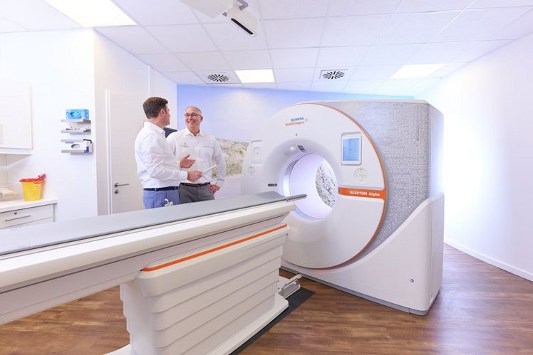
Computed Tomography (CT)

Method
In Germany, Computed Tomography may only be performed by radiologists.
Unlike a regular x-ray examination, CT creates not only a simple silhouette, but also a cross-sectional image of the corresponding organ or body section. One or two x-ray sources rotate around the affected body part during recording, while the opposite x-ray detectors collect the weakened beams left after having gone through body structures (organs, bones, soft tissue).
Converted into digital data, this information provides a slide by slide picture of an anatomical cross-section that can be reconstructed and viewed on the screen.
This Method offers further Sub-Methods
Method
In exceptional cases when MR Angiography cannot be performed, for example due to a pacemaker, we also offer CT Angiography. This, however, is carried out by using an iodinated contrast agent and x-rays. But the contrast agent is only injected into your arm vein and no catheter is inserted into the groin. Therefore, the CT Angiography procedure is performed as quickly as the MR Angiography. It does not require any follow-up, so you can take up your usual activities after examination. In general, this examination does not differ a lot from the MR Angiography.
Method
CT Coronary Angiography / Cardiac CT
The CT Coronary Angiography is performed in order to rule out narrowing of the coronary vessels in case of relevant risk factors or by abnormal symptoms. In addition, the patency of stents or of coronary artery bypass grafts can be securely detected. With the help of CT Coronary Angiography, the coronary arteries are depicted without having to insert a catheter into the groin.
Key facts about the radiation dose of CT Coronary Angiography
As in the case of cardiac catheterization, x-rays are used during CT Coronary Angiography, i.e. the examination is associated with a radiation dose. The radiation dose varies depending on the problem. In many cases, a dose used during CT Coronary Angiography can be below 1 mSv. This radiation dose is usually lower than the natural environmental radiation exposure. This consists mainly of the natural background radiation as well as of radiation produced by construction materials, and makes up an average dose of about 2.5 mSv per year. In some cases - for example when travelling by plane - it may even be significantly higher.
Who is the examination for?
- For patients with so-called „moderate, possibly even lower pre-test likelihood“ of developing coronary artery disease. Please contact us to find out whether you need a CT Angiography.
- For patients undergoing coronary artery bypass surgery. In order to evaluate the condition of the bypass vessels, CT Angiography is now a good alternative to a cardiac catheterization.
- For patients for whom the patency of a coronary artery must be verified after implantation of stents.
Advantages of CT Coronary Angiography
- In contrast to cardiac catheterization, no catheter is inserted into the artery.
- The examination is performed significantly faster than the cardiac catheterization.
- „Soft plaques“ that have not yet led to narrowing of the coronary arteries can be detected only with CT Coronary Angiography.
We perform this examination with the help of a dual-source multi-slice CT (two-tube system). The most advanced equipment is available at the Radiologie am Rathausmarkt - the Siemens Force. The Force has two flash x-ray tubes that rotate simultaneously around the patient’s body, achieving the highest temporal resolution with the lowest dose.
Method
The calcification of the coronary arteries can be detected with the help of CT Coronary Calcium Scoring. This is a sign of cardiovascular disease, since there is no lime in healthy heart vessels. With the help of computer-assisted analysis and on the basis of standardized criteria, we can evaluate whether the coronary vessels are calcified and if so, to which extent.
Based on compared values gained from a large group of patients, the risk of the presence of coronary artery disease can be estimated depending on sex and age. If there are no calcifications, coronary artery disease can be ruled out with a probability of 95 percent. If the examination shows a great amount of coronary calcium, there is a possibility that coronary artery disease is present. In this case further cardiological diagnostics are required. In the long term, the disease’s progress can be observed with the help of CT Coronary Calcium Scoring and the success of corresponding therapeutic interventions can be ensured.
Who is the examination for?
For patients who do not have typical symptoms, but who have risk factors such as heart attacks in their family history, diabetes, obesity or smoking, even if a cardiac catheterization has not been indicated yet.
Advantages of CT Coronary Calcium Scoring
- The examination is painless and takes only a few minutes.
- No contrast material is required.
- The disease’s progress and the success of the relevant therapeutic interventions can be observed in the long-term.
Key facts about the radiation dose of CT Coronary Calcium Scoring
X-rays are used during the CT Coronary Calcium Scoring. The resulting radiation dose is significantly lower than 1 mSv. Therefore, the radiation exposure is significantly lower than that from the environment which everyone is exposed to over years. The latter consists mainly of natural background radiation and radiation produced by construction materials and amounts to about 2.5 mSv per year on average. In particular cases - for example when travelling by plane - it may even be significantly higher.
We perform this examination with the help of a dual-source multi-slice CT (two-tube system). The most up-to-date device, the Siemens Force, is available at the Radiologie am Rathausmarkt. The Force has two x-ray tubes that rotate simultaneously around the patient’s body, achieving the highest temporal resolution with the lowest dose
Method
Nowadays, lung cancer can be diagnosed reliably and in a very gentle way, using so-called low-dose computed tomography. With the help of modern technology, tumours with a diameter of only a few millimetres can be detected.
The process is very suitable as an early recognition method for patients with an increased risk of lung cancer (e.g. heavy smokers). Initial studies show that many very small (< 20 mm), lung tumours can be diagnosed, which are easily operable and therefore curable.
In addition to its use as an early detection procedure for lung cancer, depending on the question to be answered and the patient’s physique, we also use low dose CT of the lung to clarify illnesses which affect the lungs only (i.e. asbestosis, sarcoidosis), and to look for tumours in high-risk patients.
Advantages and disadvantages of lung cancer early recognition with computed tomography
- The low-dose CT in multilayer technology is considered to be the most sensitive method for early recognition of lung cancer.
- The low-dose CT increases the chances of recovery since lung tumours can be detected in very early stages.
- With the help of low-dose CT, lung cancer tumours that „hide“ behind vessels and diaphragm domes can also be recognized. The conventional methods do not detect these tumours.
- With the help of computer-assisted analysis of the CT data we perform as a standard measure, the size of a tumour can be accurately measured and the smallest changes can be detected. By means of a CT follow-up examination at three to six month intervals, benign tissue changes can be differentiated from malignant ones based on a lack of growth.
- The introduction of new detector systems makes it possible to perform lung examinations with a very low radiation dose. With the multi-slice CT technology, the radiation exposure is approximately 0.2 to 0.6 mSv. By way of comparison: every citizen in Germany is, on average, exposed to a radiation dose of about 2.5 mSv from the environment every year.
Method
The virtual colonoscopy, also known as CT Colonography, is used to look at the inside of the colon and examine it for changes. Colon polyps and tumours starting from 8 mm in size can be detected early with the help of this procedure.
During the Virtual Colonoscopy no endoscope has to be inserted into the colon. The „journey“ through the colon is simulated on a computer monitor.
First of all, detailed two-dimensional images are created with the help of computed tomography. These virtual cross-sectional images are then converted into a three dimensional view of the intestine by a special computer program which allows the physician to make a virtual tour of the entire large intestine. The examination is therefore much more comfortable than conventional colonoscopy; it is painless and does not require sedation.
According to previous findings, the method is as reliable as the conventional colonoscopy when detecting polyps or bowel cancer in sizes of more than eight millimetres. A disadvantage is that the doctor cannot take a tissue sample (biopsy) during the examination. If there are suspicious changes in the intestine, a regular colonoscopy should be performed additionally in each case. However, if no pathological changes were found during the virtual colonoscopy, no further action is required.
This method is suitable for patients who cannot be examined by conventional colonoscopy due to previous illnesses or other findings.
Method
The Dual-Energy-CT requires the application of a dual source multilayer CT (two tube system). At the Radiologie am Rathausmarkt, the most up-to-date device, the Definition Flash of Siemens, is available.
The Definition Flash has two x-ray tubes that rotate simultaneously around the patient’s body.
By using different tube voltages (dual-energy), calcifications and calcified changes can be detected precisely (gout) or stones consisting purely of uric acid can be differentiated from other or mixed stones (kidney stones). Administering contrast agents is not necessary.
Method
The focus of the practice is on musculoskeletal imaging in order to diagnose acute and chronic illnesses of the whole musculoskeletal system in adults and children. Cartilage is of central significance for the integrity of the joints. Using ultra-high-resolution MRT, an unparalleled image can be created of the deterioration of cartilage – even in its early stages. An exact diagnosis forms the basis for early and efficient treatment. Of course, we also offer measurement of bone density for the early recognition or progress and therapy monitoring of osteoporosis.