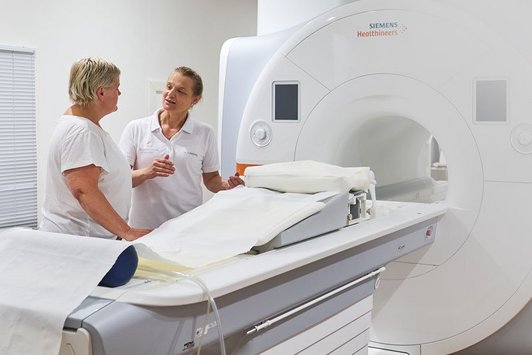
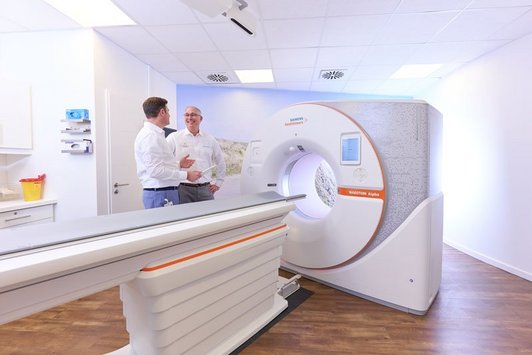
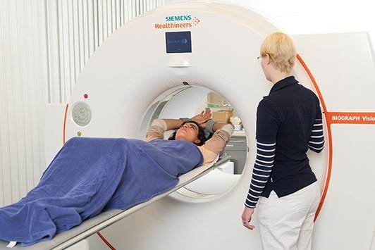
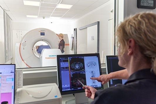
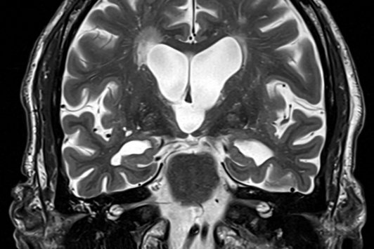
Methods

Diagnostic Procedures

  • Magnetic Resonance Imaging (MRI)
    • This Method offers further Sub-Methods:

    • MR Angiography

      This method makes it possible to examine your blood vessels without surgical intervention. With the help of the magnetic field technique, we create a three-dimensional model of your vessels so we can see if everything is running “smoothly” there. It helps to recognize narrowing and tissue weaknesses (aneurysms) that may cause circulation problems, stroke or bleeding. The MR Angiography is so advanced that its image quality is the same as that of conventional angiography. In case of specific problems beyond vasoconstriction, e.g. in an upper or lower limb artery, MR Angiography provides more precise information than conventional angiography does. 

      Advantages of MR Angiography compared with X-ray Angiography (DSA)

      The standard method of examination used to be X-ray Angiography. For this purpose, a thin tube (catheter) was inserted from the groin into the artery. While x-ray contrast media was injected through the catheter under high pressure, we would create x-ray images which depicted the blood vessel filled with contrast agent.

      MR Angiography no longer requires inserting a thin tube (catheter) from the groin into the artery. This saves approximately four hours of semi-hospitalization. In MR Angiography a contrast agent is injected into the vein. In addition to that, no x-rays are used in MRAngiography, but the vessels are visualized with the help of a strong magnetic field.

      Advantages of MR Vessel examination

      • In MR Angiography, no catheter is inserted into the femoral artery. The contrast material is injected only into the arm vein.
      • In the case of specific problems, MR Angiography provides more detailed information than the regular x-ray angiography.
      • The examination is done quickly and without semi-hospitalization.
      • There are no problems such as bleeding or clotting disorders.
      • MR Angiography does not use x-rays. The vessels are shown with the help of a strong magnetic field.
      • The contrast agent used in MR Angiography does not contain iodine and can therefore be used for patients suffering from hyperthyroidism.

      The procedure is completed without x-rays. However, in Germany it may only be performed by radiologists.

      Practices

      MR Angiography
    • MR Enteroclysis (Small Intestine)

      With the help of the magnetic resonance imaging it is possible to see the small intestine. Thus, MRI is an alternative to conventional examination of the small intestine with enteroclysis. There is no need to insert a probe into the small intestine.

      MRI helps to visualize not only changes in the inner bowels, but also thickening or inflammation of the bowel wall and its environment. The examination is used to diagnose inflammatory bowel diseases such as Crohn’s disease and ulcerative colitis, the infestation and the extension of the disease.

      The procedure is performed completely without x-rays. However, in Germany it may only be performed by radiologists.

      Practices

      MR Enteroclysis Small Intestine
    • Prostate MRI

      The examination of the prostate with the help of magnetic resonance imaging (MRI) is a modern diagnostic method to combat prostate cancer. This method combines several advantages: firstly, the organ to be examined can be visually separated into different layers of tissue. Furthermore, it is non-invasive and, unlike the computer tomography, x-rays will not be used to create images. The examination of the prostate is performed with the help of two coils. The painless insertion of the endorectal coil into the rectum makes it possible for the probe to approach the prostate and thus create high-resolution imaging.

      The procedure is performed completely without x-rays. However, in Germany it may only be performed by radiologists.

      Who is this examination for?

      • For men who have an increased PSA level 
      • For men whose tissue sample taken after a PSA increase was benign, but their PSA level continued to increase (MRI prostate biopsy before second punch biopsy)
      • As additional guidance for the upcoming decision on treatment (e. g. surgery, radiotherapy, hormonal therapy?) for men with histologically diagnosed prostate cancer.
      • When, after removing the prostate, the PSA level rises again. Is this a local recurrence? Is there a lymph node enlargement in the pelvic area? Many radiation therapy institutions require this cross-sectional imaging procedure before the start of therapy.

      Advantages and disadvantages of Prostate MRI

      • No x-rays are required.
      • A tumour, and in particular the question of the size and advancement of a prostate cancer in its surrounding. The infiltration of the seminal vesicles is safer with MRI than with other cross-sectional imaging methods.
      • The lymph nodes of the pelvis and especially the regional lymph nodes in the immediate vicinity of the prostate gland can be detected with high resolution on three levels with the endorectal coil. 

      Disadvantage: In the past, severe inflammation of the prostate (prostatitis) could be similar to morphologic carcinoma and a tumour.

      Practices

      Prostate MRI
    • Pelvic MRI

      Magnetic resonance imaging (MRI) of the pelvis - also called nuclear resonance imaging of the pelvis - is a radiological examination method used to visualize structures in the area of the pelvis with the help of a magnetic field.

      The Pelvic MRI is a very accurate diagnostic procedure used in case of the following diseases and symptoms:

      • Suspected disorders in the pelvic floor such as defecation disorders
      • Suspected prolapse of the uterus or bladder due to muscular weakness of the pelvic floor muscles
      • Tumours in the pelvic floor area
      • Congenital defects in the pelvic floor area

      The procedure is performed completely without x-rays. However, in Germany it may only be performed by radiologists.

      Practices

      Pelvic MRI
    • MR Mammography

      Sometimes, the MRI examination of the breast can be used not only complementarily, but also as an alternative to x-ray mammography. It can help to clarify certain issues that may have possibly been left open after mammography and other examination procedures of the breast. As no x-rays are used during magnetic resonance imaging (MRI), this method is considered to be especially gentle.

      The breast MRI is performed, for example, in order to precisely examine patients with breast cancer before surgery. In the after-care of patients who have already undergone breast surgery or have breast implants, MR Mammography is also used. This method complements the conventional x-ray mammography procedure, especially for women with dense tissue structures. 

      MR Mammography indisputably has the highest sensitivity and preciseness for the detection of breast tumours that are already growing invasively.

      Who are the examinations intended for?

      • For women with diagnosed changes in the breast tissue (such as a digital rectal examination, or abnormalities in the x-ray or ultrasound examination): Magnetic Resonance Imaging of the breast may clarify whether the changes in the breast are benign or malignant, in the cases when the usual screening tests such as mammography (x-ray) and ultrasound do not provide reliable information.

      • For patients with breast cancer, as a test before surgery to exclude other focuses of cancer and to plan the operative procedure. Should chemotherapy be required before surgery, its efficiency and progress can be checked. 
      • For breast cancer patients in after-care to exclude a scar recurrence (tumour formation on the scar) with greater safety.
      • For women with breast implants (e.g. silicone): The implants can interfere with the conventional diagnostic mammography (the implant covers large parts of the glandular tissue as a ‘shadow’). In addition, the position and integrity of the implants can be checked. 
      • For patients who may have specific reasons which do not allow them to undergo breast cancer screening or x-ray mammography or they do not wish to do it: For example, young women with frequent breast cancer cases in the family history, who need an early detection of breast cancer as early as after the age of 30.

      Advantages and disadvantages of magnetic resonance imaging of the female breast

      • The images are created with the help of a strong magnetic field. X-rays are not required.
      • The magnetic resonance imaging can be so good that it complements mammography and / or breast ultrasound in case of appropriate indications.

      Magnetic resonance imaging alone cannot always distinguish benign tumours from malignant ones or detect breast cancer in its preliminary stages (carcinoma in situ).

      The procedure is performed completely without x-rays. However, in Germany it may only be performed by radiologists. 

      Practices

      MR Mammography
    • Cardiac MRI

      Thanks to rapid technological and scientific development, the magnetic resonance imaging of the heart - Cardiac MRI for short, is now an established method for the diagnosis of various heart diseases. The Cardiac MRI is an accurate method with which to measure heart size and function. In addition to that, even the smallest change in cardiac output can be reliably detected by MRI.

      Coronary artery disease (CAD) – initial diagnosis due to detection of vascular disorders

      CAD is one of the most common cardiac diseases. It is caused by atherosclerosis or „narrowing“ of the coronary vessels, leading to vascular disorders of the heart muscle.

      In later stages it can often cause some common symptoms such as angina pectoris or heart attack. With the help of MRI, it is possible to detect blood flow disorders of the heart muscle and measure the heart size and function. In addition to that, so-called „silent“ heart attacks are detected, which the patient did not notice. They can be the only signs of CHD.

      Coronary artery disease - detection and assessment of heart attack

      Despite the progress in diagnostics, a sudden heart attack remains a constant threat. After a heart attack, the heart might only work in a limited manner. A Cardiac MRI can precisely determine cardiac function, size, the nature and prognosis of heart attack. It is considered to be the most accurate method for detecting scar tissue in the heart muscle.

      The MRI of the heart is also used for diagnosing other heart diseases:

      • Congenital heart defects in childhood and adulthood
      • Acquired valve defect
      • Heart muscle disease (cardiomyopathy)
      • Heart muscle inflammation (myocarditis)
      • Thrombi or tumours in the heart.

      Advantages of magnetic resonance imaging of the heart

      The MRI of the heart is also used for diagnosing other heart diseases:

      • MRI of the heart can precisely determine cardiac function, size, as well as the nature and prognosis of heart attack.
      • It is considered to be the most accurate method for detecting scar tissue in the heart muscle, or so-called „silent“ heart attacks.
      • No heart catheters need to be inserted into the artery.

      Practices

      Cardiac MRI
    • Whole Body Imaging (MRI), Early Diagnosis & Prevention

      Whole body MRT makes it possible for us to view an image of the whole body – all organs, the skeleton, the spine, the circulatory system and the joints. The examination is principally for the early detection of cancer. Depending on the patient’s age and genetic or other previous conditions, other questions can be answered – such as whether there is a narrowing of the blood vessels. The examination can take place without x-ray and usually without the use of contrast agent.

      Practices

      Whole Body Imaging MRI Early Diagnosis Prevention
    • Sports Medicinal & Musculoskeletal Diagnostic Procedures

      The focus of the practice is on musculoskeletal imaging in order to diagnose acute and chronic illnesses of the whole musculoskeletal system in adults and children. Cartilage is of central significance for the integrity of the joints. Using ultra-high-resolution MRT, an unparalleled image can be created of the deterioration of cartilage – even in its early stages. An exact diagnosis forms the basis for early and efficient treatment. Of course, we also offer measurement of bone density for the early recognition or progress and therapy monitoring of osteoporosis.

      Practices

      Sports Medicinal Musculoskeletal Diagnostic Procedures

    Magnetic Resonance Imaging (MRI)

    Magnetic Resonance Imaging (MRI) is a medical imaging technique used primarily in medical diagnostics to visualize internal structure, the function of tissues and body organs.

    MRI uses electric and magnetic fields. The high-frequency pulses (radio waves) stimulate atoms with an odd mass number (dipoles) in the human body, so that they vibrate. Different types of tissues of the body are determined by different so-called relaxation times. In this way, the contrasts between organs and tissues are made visible.

    Images showing the body on each possible level depict all organs in detail, thus meaning that potential pathological changes can be recognized.

    The process is performed completely without x-rays. However, in Germany it may only be used by radiologists.

    Practices

    Magnetic Resonance Imaging MRI
  • Computed Tomography (CT)
    • This Method offers further Sub-Methods:

    • CT Angiography

      In exceptional cases when MR Angiography cannot be performed, for example due to a pacemaker, we also offer CT Angiography. This, however, is carried out by using an iodinated contrast agent and x-rays. But the contrast agent is only injected into your arm vein and no catheter is inserted into the groin. Therefore, the CT Angiography procedure is performed as quickly as the MR Angiography. It does not require any follow-up, so you can take up your usual activities after examination. In general, this examination does not differ a lot from the MR Angiography.

      Practices

      CT Angiography
    • CT Coronary-Angiography/Cardiac CT

      CT Coronary Angiography / Cardiac CT

      The CT Coronary Angiography is performed in order to rule out narrowing of the coronary vessels in case of relevant risk factors or by abnormal symptoms. In addition, the patency of stents or of coronary artery bypass grafts can be securely detected. With the help of CT Coronary Angiography, the coronary arteries are depicted without having to insert a catheter into the groin.

      Key facts about the radiation dose of CT Coronary Angiography

      As in the case of cardiac catheterization, x-rays are used during CT Coronary Angiography, i.e. the examination is associated with a radiation dose. The radiation dose varies depending on the problem. In many cases, a dose used during CT Coronary Angiography can be below 1 mSv. This radiation dose is usually lower than the natural environmental radiation exposure. This consists mainly of the natural background radiation as well as of radiation produced by construction materials, and makes up an average dose of about 2.5 mSv per year. In some cases - for example when travelling by plane - it may even be significantly higher.

      Who is the examination for?

      • For patients with so-called „moderate, possibly even lower pre-test likelihood“ of developing coronary artery disease. Please contact us to find out whether you need a CT Angiography.
      • For patients undergoing coronary artery bypass surgery. In order to evaluate the condition of the bypass vessels, CT Angiography is now a good alternative to a cardiac catheterization.
      • For patients for whom the patency of a coronary artery must be verified after implantation of stents.

      Advantages of CT Coronary Angiography

      • In contrast to cardiac catheterization, no catheter is inserted into the artery.
      • The examination is performed significantly faster than the cardiac catheterization.
      • „Soft plaques“ that have not yet led to narrowing of the coronary arteries can be detected only with CT Coronary Angiography.

      We perform this examination with the help of a dual-source multi-slice CT (two-tube system). The most advanced equipment is available at the Radiologie am Rathausmarkt - the Siemens Force. The Force has two flash x-ray tubes that rotate simultaneously around the patient’s body, achieving the highest temporal resolution with the lowest dose.

      Practices

      CT Coronary-Angiography Cardiac CT
    • CT Coronary Calcium Scoring

      The calcification of the coronary arteries can be detected with the help of CT Coronary Calcium Scoring. This is a sign of cardiovascular disease, since there is no lime in healthy heart vessels. With the help of computer-assisted analysis and on the basis of standardized criteria, we can evaluate whether the coronary vessels are calcified and if so, to which extent.

      Based on compared values gained from a large group of patients, the risk of the presence of coronary artery disease can be estimated depending on sex and age. If there are no calcifications, coronary artery disease can be ruled out with a probability of 95 percent. If the examination shows a great amount of coronary calcium, there is a possibility that coronary artery disease is present. In this case further cardiological diagnostics are required. In the long term, the disease’s progress can be observed with the help of CT Coronary Calcium Scoring and the success of corresponding therapeutic interventions can be ensured.

      Who is the examination for?

      For patients who do not have typical symptoms, but who have risk factors such as heart attacks in their family history, diabetes, obesity or smoking, even if a cardiac catheterization has not been indicated yet.

      Advantages of CT Coronary Calcium Scoring

      • The examination is painless and takes only a few minutes.
      • No contrast material is required.
      • The disease’s progress and the success of the relevant therapeutic interventions can be observed in the long-term.

      Key facts about the radiation dose of CT Coronary Calcium Scoring

      X-rays are used during the CT Coronary Calcium Scoring. The resulting radiation dose is significantly lower than 1 mSv. Therefore, the radiation exposure is significantly lower than that from the environment which everyone is exposed to over years. The latter consists mainly of natural background radiation and radiation produced by construction materials and amounts to about 2.5 mSv per year on average. In particular cases - for example when travelling by plane - it may even be significantly higher.

      We perform this examination with the help of a dual-source multi-slice CT (two-tube system). The most up-to-date device, the Siemens Force, is available at the Radiologie am Rathausmarkt. The Force has two x-ray tubes that rotate simultaneously around the patient’s body, achieving the highest temporal resolution with the lowest dose

      Practices

      CT Coronary Calcium Scoring
    • Low-dose CT of the Lung

      Nowadays, lung cancer can be diagnosed reliably and in a very gentle way, using so-called low-dose computed tomography. With the help of modern technology, tumours with a diameter of only a few millimetres can be detected.

      The process is very suitable as an early recognition method for patients with an increased risk of lung cancer (e.g. heavy smokers). Initial studies show that many very small (< 20 mm), lung tumours can be diagnosed, which are easily operable and therefore curable.

      In addition to its use as an early detection procedure for lung cancer, depending on the question to be answered and the patient’s physique, we also use low dose CT of the lung to clarify illnesses which affect the lungs only (i.e. asbestosis, sarcoidosis), and to look for tumours in high-risk patients.

      Advantages and disadvantages of lung cancer early recognition with computed tomography

      • The low-dose CT in multilayer technology is considered to be the most sensitive method for early recognition of lung cancer.
      • The low-dose CT increases the chances of recovery since lung tumours can be detected in very early stages.
      • With the help of low-dose CT, lung cancer tumours that „hide“ behind vessels and diaphragm domes can also be recognized. The conventional methods do not detect these tumours.
      • With the help of computer-assisted analysis of the CT data we perform as a standard measure, the size of a tumour can be accurately measured and the smallest changes can be detected. By means of a CT follow-up examination at three to six month intervals, benign tissue changes can be differentiated from malignant ones based on a lack of growth.
      • The introduction of new detector systems makes it possible to perform lung examinations with a very low radiation dose. With the multi-slice CT technology, the radiation exposure is approximately 0.2 to 0.6 mSv. By way of comparison: every citizen in Germany is, on average, exposed to a radiation dose of about 2.5 mSv from the environment every year.

      Practices

      Low-dose CT of the Lung
    • Virtual CT Colonoscopy

      The virtual colonoscopy, also known as CT Colonography, is used to look at the inside of the colon and examine it for changes. Colon polyps and tumours starting from 8 mm in size can be detected early with the help of this procedure.

      During the Virtual Colonoscopy no endoscope has to be inserted into the colon. The „journey“ through the colon is simulated on a computer monitor.

      First of all, detailed two-dimensional images are created with the help of computed tomography. These virtual cross-sectional images are then converted into a three dimensional view of the intestine by a special computer program which allows the physician to make a virtual tour of the entire large intestine. The examination is therefore much more comfortable than conventional colonoscopy; it is painless and does not require sedation.

      According to previous findings, the method is as reliable as the conventional colonoscopy when detecting polyps or bowel cancer in sizes of more than eight millimetres. A disadvantage is that the doctor cannot take a tissue sample (biopsy) during the examination. If there are suspicious changes in the intestine, a regular colonoscopy should be performed additionally in each case. However, if no pathological changes were found during the virtual colonoscopy, no further action is required.

      This method is suitable for patients who cannot be examined by conventional colonoscopy due to previous illnesses or other findings.

      Practices

      Virtual CT Colonoscopy
    • Dual-Energy CT (Gout)

      The Dual-Energy-CT requires the application of a dual source multilayer CT (two tube system). At the Radiologie am Rathausmarkt, the most up-to-date device, the Definition Flash of Siemens, is available.

      The Definition Flash has two x-ray tubes that rotate simultaneously around the patient’s body. 

      By using different tube voltages (dual-energy), calcifications and calcified changes can be detected precisely (gout) or stones consisting purely of uric acid can be differentiated from other or mixed stones (kidney stones). Administering contrast agents is not necessary.

      Practices

      Dual-Energy CT Gout
    • Dual-Energy CT (Kidney)

      The Dual-Energy-CT requires the application of a dual source multilayer CT (two tube system). At the Radiologie am Rathausmarkt, the most up-to-date device, the Definition Flash of Siemens, is available.

      The Definition Flash has two x-ray tubes that rotate simultaneously around the patient’s body. 

      By using different tube voltages (dual-energy), calcifications and calcified changes can be detected precisely (gout) or stones consisting purely of uric acid can be differentiated from other or mixed stones (kidney stones). Administering contrast agents is not necessary.

      Practices

      Dual-Energy CT Kidney
    • Sports Medicinal & Musculoskeletal Diagnostic Procedures

      The focus of the practice is on musculoskeletal imaging in order to diagnose acute and chronic illnesses of the whole musculoskeletal system in adults and children. Cartilage is of central significance for the integrity of the joints. Using ultra-high-resolution MRT, an unparalleled image can be created of the deterioration of cartilage – even in its early stages. An exact diagnosis forms the basis for early and efficient treatment. Of course, we also offer measurement of bone density for the early recognition or progress and therapy monitoring of osteoporosis.

      Practices

      Sports Medicinal Musculoskeletal Diagnostic Procedures

    Computed Tomography (CT)

    In Germany, Computed Tomography may only be performed by radiologists.

    Unlike a regular x-ray examination, CT creates not only a simple silhouette, but also a cross-sectional image of the corresponding organ or body section. One or two x-ray sources rotate around the affected body part during recording, while the opposite x-ray detectors collect the weakened beams left after having gone through body structures (organs, bones, soft tissue).

    Converted into digital data, this information provides a slide by slide picture of an anatomical cross-section that can be reconstructed and viewed on the screen.

    Practices

    Computed Tomography CT
  • Mamma Diagnostics
    • This Method offers further Sub-Methods:

    • MR Mammography

      Sometimes, the MRI examination of the breast can be used not only complementarily, but also as an alternative to x-ray mammography. It can help to clarify certain issues that may have possibly been left open after mammography and other examination procedures of the breast. As no x-rays are used during magnetic resonance imaging (MRI), this method is considered to be especially gentle.

      The breast MRI is performed, for example, in order to precisely examine patients with breast cancer before surgery. In the after-care of patients who have already undergone breast surgery or have breast implants, MR Mammography is also used. This method complements the conventional x-ray mammography procedure, especially for women with dense tissue structures. 

      MR Mammography indisputably has the highest sensitivity and preciseness for the detection of breast tumours that are already growing invasively.

      Who are the examinations intended for?

      • For women with diagnosed changes in the breast tissue (such as a digital rectal examination, or abnormalities in the x-ray or ultrasound examination): Magnetic Resonance Imaging of the breast may clarify whether the changes in the breast are benign or malignant, in the cases when the usual screening tests such as mammography (x-ray) and ultrasound do not provide reliable information.

      • For patients with breast cancer, as a test before surgery to exclude other focuses of cancer and to plan the operative procedure. Should chemotherapy be required before surgery, its efficiency and progress can be checked. 
      • For breast cancer patients in after-care to exclude a scar recurrence (tumour formation on the scar) with greater safety.
      • For women with breast implants (e.g. silicone): The implants can interfere with the conventional diagnostic mammography (the implant covers large parts of the glandular tissue as a ‘shadow’). In addition, the position and integrity of the implants can be checked. 
      • For patients who may have specific reasons which do not allow them to undergo breast cancer screening or x-ray mammography or they do not wish to do it: For example, young women with frequent breast cancer cases in the family history, who need an early detection of breast cancer as early as after the age of 30.

      Advantages and disadvantages of magnetic resonance imaging of the female breast

      • The images are created with the help of a strong magnetic field. X-rays are not required.
      • The magnetic resonance imaging can be so good that it complements mammography and / or breast ultrasound in case of appropriate indications.

      Magnetic resonance imaging alone cannot always distinguish benign tumours from malignant ones or detect breast cancer in its preliminary stages (carcinoma in situ).

      The procedure is performed completely without x-rays. However, in Germany it may only be performed by radiologists. 

      Practices

      MR Mammography
    • Low-dose Mammography and Tomosynthesis

      The Radiologische Allianz also offers Low Dose Mammography, Tomosynthesis (3D mammography of the breast) and often a Computer Assisted second Diagnosis, (CAD).

      Low Dose Mammography

      A mammogram is an examination of the breast using x-ray. It is used to diagnose breast cancer in its early stages, and in connection with breast cancer operations. A mammogram can make the tiniest tumours visible at very early stages when they cannot yet be felt, thus improving the patient’s chances of recovery. The Radiologische Allianz works with digital mammograms as its standard method.

      Scientific studies confirm that mammograms are the best way to diagnose tiny tumours in their early stages. For this reason, this examination method forms the basis of the Radiologische Allianz’s breast cancer prevention approach.

      With the same image quality, the low dose mammogram makes it possible to reduce the radiation dose by up to 30 percent by dispensing with the anti-scatter grid. In view of the large number of mainly healthy women who are examined as part of early detection mammography, this is a significant change.

      Tomosynthesis

      In 3D tomosynthesis, three-dimensional images of the breast are created. This means that we can recognise tumours which are hidden by overlapping tissue – something which was not possible until now. The presentation of the breast tissue as a cross-section without anything superimposed on it makes a more precise diagnosis possible, and reduces the number of so-called false positive results. This technique is used by the Radiologische Allianz in addition to the usual digital mammograpy and other breast diagnostics methods, in particular for patients with dense mammary tissue and an inconclusive diagnosis.

      Practices

      Low-dose Mammography and Tomosynthesis
    • Tomosynthesis

      The Radiologische Allianz also offers Low Dose Mammography, Tomosynthesis (3D mammography of the breast) and often a Computer Assisted second Diagnosis, (CAD).

      Tomosynthesis

      In 3D tomosynthesis, three-dimensional images of the breast are created. This means that we can recognise tumours which are hidden by overlapping tissue – something which was not possible until now. The presentation of the breast tissue as a cross-section without anything superimposed on it makes a more precise diagnosis possible, and reduces the number of so-called false positive results. This technique is used by the Radiologische Allianz in addition to the usual digital mammograpy and other breast diagnostics methods, in particular for patients with dense mammary tissue and an inconclusive diagnosis.

      Low Dose Mammography

      A mammogram is an examination of the breast using x-ray. It is used to diagnose breast cancer in its early stages, and in connection with breast cancer operations. A mammogram can make the tiniest tumours visible at very early stages when they cannot yet be felt, thus improving the patient’s chances of recovery. The Radiologische Allianz works with digital mammograms as its standard method.

      Scientific studies confirm that mammograms are the best way to diagnose tiny tumours in their early stages. For this reason, this examination method forms the basis of the Radiologische Allianz’s breast cancer prevention approach.

      With the same image quality, the low dose mammogram makes it possible to reduce the radiation dose by up to 30 percent by dispensing with the anti-scatter grid. In view of the large number of mainly healthy women who are examined as part of early detection mammography, this is a significant change.

      Practices

      Tomosynthesis

    Mamma Diagnostics

    The Radiologische Allianz offers the whole range of breast diagnostics along with the newest equipment. In addition to digital mammograms, we also use the following diagnostic procedures:

    Tomosynthesis

    In 3D tomosynthesis, three-dimensional images of the breast are created. This means that we can recognise tumours which are hidden by overlapping tissue – something which was not possible until now. The presentation of the breast tissue without anything superimposed on it makes a more precise diagnosis possible, and reduces the number of so-called false positive results.

    Sonography

    Sonography is an additional procedure which can be used to characterise results which cannot be evaluated reliably using a mammogram alone, and to confirm results found by touch. For women aged 40 or younger, the imaging procedure is the first choice. Women over 40 should also have a mammogram if they have high mammary gland density.

    Biopsy as outpatient procedure (vacuum-assisted and punch biopsies)

    Inconclusive results can be clarified by taking tissue samples under local anaesthetic. The removal of the tissue is monitored using an imaging procedure.

    MR Mammography

    During MRI of the breast, images are created using contrast agent. This is the most sensitive procedure available for diagnosing breast cancer. It can be used as an additional method in the case of inconclusive results, and to answer specific questions as part of post-operative aftercare.

    Practices

    Mamma Diagnostics
  • Nuclear Medicine/SPECT/Scintigraphy
    • This Method offers further Sub-Methods:

    • Scintigraphy

      Nuclear medicine has existed for many decades. It is an important component of diagnostics and therapy. 

      The principle is always the same: we know certain metabolic pathways within and between the organs. Low level radiating substances (radiopharmaceuticals) are connected to substances that „go“ this way. A gamma camera detects these substances and a computer converts the information into images. At the same time, the temporal change and spatial distribution of the radiopharmaceutical in a certain area or in the whole body are recorded: that is how a scintigraphy is made. In this way, an abnormal metabolic process can be regionally allocated.

      We use modern gamma cameras for nuclear medical examinations. They help to answer all questions posed to nuclear medical diagnostics. Gamma cameras can electronically register the temporal and spatial distribution of the gamma radiation emitted from the patient and send them to a data processing system. This system can process the data appearing in very large quantities within a very short period of time, visualize it and save it. In addition to this, SPECT images (Single Photon Emission Computed Tomography – nuclear medical sliced images) of a very high quality can be developed.

      Szintigraphie und SPECT/CT der Nebenschilddrüse

      Practices

      Scintigraphy
    • Radiosynoviorthese (RSO)

      Radiosynoviorthese means restoration (orthesis) of the joint mucosa (synovialis) with the help of radioactive isotopes. It is a very effective method, applied in the case of painful inflammatory changes in the joints. This method has existed for two decades. A radioactive substance is injected into a diseased joint. After the injection into the joint, it distributes itself homogenously in the joint fluid. The substance is absorbed by the surface cells of the diseased joint mucosa and produces an anti-inflammatory effect. Gradually, a sloughing of the mucous surface occurs, and the mucosa swelling decreases. The finest canals from which the fluid can penetrate into the joint are closed, and the finest pain sensors are turned off. This way the pain is decreased or eliminated, and the function of the joint is improved. The effect occurs gradually, sometimes after several days, in some cases after weeks or months. The final treatment effect can be evaluated after about 6 months. 

      Advantages of Radiosynoviorthese:

      The actual therapy is a small intervention which can be carried out on an outpatient basis, though it has been possible only for the past few years. Rehabilitation, the way it was necessary after a traditional surgery, is no longer needed. The therapy can also be carried out on patients with an increased surgical risk. Many joints can be treated simultaneously or at short intervals. If the effect is low, e.g. if the pathological changes of joints have already advanced a lot, the therapy can be repeated. Normally it is enough to carry out one treatment on a joint.

      Areas of use

      The focus is on chronic polyarthritis (inflammatory rheumatism of the joints). This method is used, among other things, to treat psoriatic arthritis, active arthrosis – of the knees, for example (osteoarthritis) and for so-called irritable knee and active finger polyarthrosis.

      Rheumatic complaints can also be treated using RSO. Rheumatism is an illness which affects the entire body, but its cause is largely unknown. It is an autoimmune disease – this means that the body reacts aggressively with inflammation towards parts of the person’s own body, because it recognises these wrongly as being foreign substances. This inner battle leads in particular to damage in the joints, and within them to inflammation of the joint mucous membranes (synovialitis). The joint mucous membrane often proliferates like coral, destroys the cartilage and forces its way into the bones, where it can tear ligaments and tendons, and damage or destroy joints at different speeds. In addition to treatment with medicine, joint operations are often necessary too. If one or a few joints are particularly affected, it is recommended to use radiosynoviorthese as early as possible as a low-intensity radiation therapy to prevent inflammation – if possible, before serious damage is caused to the joints.

      Practices

      Radiosynoviorthese RSO

    Nuclear Medicine/SPECT/Scintigraphy

    Nuclear medicine has existed for many decades. It is an important component of diagnostics and therapy. 

    The principle is always the same: we know certain metabolic pathways within and between the organs. Low level radiating substances (radiopharmaceuticals) are connected to substances that „go“ this way. A gamma camera detects these substances and a computer converts the information into images. At the same time, the temporal change and spatial distribution of the radiopharmaceutical in a certain area or in the whole body are recorded: that is how a scintigraphy is made. In this way, an abnormal metabolic process can be regionally allocated.

    We use modern gamma cameras for nuclear medical examinations. They help to answer all questions posed to nuclear medical diagnostics. Gamma cameras can electronically register the temporal and spatial distribution of the gamma radiation emitted from the patient and send them to a data processing system. This system can process the data appearing in very large quantities within a very short period of time, visualize it and save it. In addition to this, SPECT images (Single Photon Emission Computed Tomography – nuclear medical sliced images) of a very high quality can be developed.

    Practices

    Nuclear Medicine SPECT Scintigraphy
  • Digital X-ray

    Digital X-ray

    X-rays are electromagnetic waves which are produced with an X-ray tube for medical purposes. In German, they are named after Wilhelm Conrad Röntgen, who discovered them in 1895 in Würzburg. In the x-ray system, the x-rays developed by it are sent through the body area being examined, and then the photons let through are transmitted to a digital detector system for the development of an image. The dose is accurately recorded and documented.

    The dose the patient is exposed to during an x-ray examination, is kept as low as possible. Compared with the early 20th century, less than 1 percent of the radiation dose needed at that time is needed today for creating an x-ray image.

    The Radiologische Allianz has switched completely to digital radiography. The advantages are the lower radiation dose and the fact that it is possible to save the image in a digital archive. We can mostly forgo usual x-ray images. Patients will receive prints and a CD, on which the data are saved instead. So the patients can look at the results of their examination on any modern PC or tablet.

    Practices

    Digital X-ray
  • PET/CT (in the PET/CT Centre of Hamburg)

    PET/CT (in the PET/CT Centre of Hamburg)

    PET/CT (Positron-Emission-Tomography and Computed Tomography)

    PET/CT – the most modern diagnostic procedure in oncology – reliably tracks down cancerous cells due to their increased metabolism, thus making it possible to diagnose cancer precisely, while it is still in its early stages.

    The PET/CT is a combination of Positron-Emission-Tomography (PET) and Computed Tomography (CT) in one device. It combines two different imaging techniques, which makes it possible to provide an exact image of the body structure and function in one and the same image. The exact location, size, activity and spread of a tumour within the body can be determined. During a single examination under the same conditions, different advantages of both methods supplement each other, creating a good image and helping to make a precise diagnosis. Studies have shown that accuracy of diagnosis based on a combined PET/CT examination results has increased compared with two scans performed separately. For you as a patient, in addition to the optimal diagnosis there is one more advantage - only one examination appointment is needed and the examination time is half that of the separate examinations. 

    PET

    PET is a nuclear medical procedure which in most cases uses the low-active radioactive substance similar to dextrose F-18 fluorodeoxyglucose (FDG). Our body cells need dextrose (glucose) as a fuel, and it accumulates in many cancer cells because they grow fast and have a significant demand for fuel. The PET camera produces a picture of the distribution of sugar within the body: the healthy tissue takes up very little glucose, so „cancer cells“ can be identified. They can be recognized early and definitely with the PET, but their exact size and position cannot be determined exactly. The data is presented in a form of cross-sectional images, full-body images and three-dimensional images.

    CT

    CT is an established x-ray procedure which uses x-rays to create finely structured layered images of the body. In some cases it is necessary to use a contrast agent in order to create clearer images. The CT can be used to precisely define the size and position of the focal point of the cancer, but it is often not able to differentiate well enough or early enough between healthy tissue and cancerous tissue.

    PET/CT and Radiotherapy

    PET/CT is an important planning tool for radiation therapy. When planning Radiotherapy based on merged data provided by PET/CT, the target volumes can be defined even more precisely. The exact localization of the radiation field makes it possible to provide individual treatment and improve the chances of recovery.

    For more information see www.petct-zentrum-hamburg.de

    Practices

    PET CT in the PET CT Centre of Hamburg
  • Neuroradiology

    Neuroradiology

    Neuroradiology is the examination and treatment of diseases of the central and peripheral nervous system with the help of imaging procedures such as Magnetic Resonance Imaging (MRI) und Computed Tomography (CT). The visualization of vascular diseases, for example a narrowing of the carotid artery, can be performed by means of MR or CT angiography instead of an invasive intra-arterial angiography. Neuroradiology is a subfield of radiology, which requires an additional specialization.

    The most frequent and important examination is the MRI of the central nervous system as well as of the spinal column and brain. In many cases, such as after a nerve root compression caused by a herniated disc, it is not necessary to use intravenous contrast agents.

    Many common clinical problems can be clarified with the help of computed tomography. In addition to this, calcifications and bone structures can be presented in great detail. CT Angiography can be a reasonable alternative to an MR Angiography when an especially high resolution is required, for example a tear of the posterior cerebral artery (vertebral dissection). Furthermore, the CT helps to precisely place an injection needle near a painful nervous root (Periradicular Therapy) or to make a contrasted visualization of the spinal canal (CT-Myelography).

    Integrated Imaging of the Nervous System

    By this, we understand the use of imaging procedures to clarify illnesses and functional disorders of the nervous system. It is less about simply showing the organ (brain, spinal cord, nerve) than about the type of disorder – modern imaging procedures can make a crucial contribution to explaining this. The procedure can be used, for example, to investigate cerebrovascular illnesses such as an impending circulatory disorder in the brain (stroke), to assess the risk of carotid artery stenosis, inflammation of the brain and spinal cord, memory disorders (dementia), illnesses of the white brain matter, tumour diseases and relapses following treatment, unclear neurological symptoms, and illnesses of peripheral nerves.

    We use specific special procedures for this purpose:

    • Blood vessel examinations (MR and CT angiographies)
    • Measurements of blood flow to the brain (perfusion measurement)
    • Plaque imaging (detailed picture of a narrowing of the carotid arteries)
    • MR spectroscopy for the differentiation of changes in tissue
    • Diffusion imaging for the recognition of brain tissue affected by circulatory disorders, cell-rich changes, and nerve pathways.
    • MR Neurography

    Practices

    Neuroradiology

Radiation Therapy

  • Radiation Therapy

    Radiation Therapy

    Radiation Therapy or Radiooncology plays an important role in the treatment of malignant diseases. Nowadays, about 45 to 50% of all people who suffer from cancer (children and adults) can be cured with the help of modern treatment procedures. 50 to 60% of those who have been cured have undergone radiotherapy as part of their treatment.

    It is carried out either as radiotherapy alone or in combination with surgical measures and/or chemotherapy. Most patients and many physicians often do not understand well enough that radiation therapy makes a significant contribution to curing cancer or is even the decisive part of treatment.

    Overmore Radiation Therapy plays an important role in alleviating the symptoms of cancer disease. Many patients who can no longer be cured benefit from pain relief or restoration of body functions (e.g. ability to swallow, ability to walk). For these patients, their disease should be considered as chronic, which requires therapy measures at various intervals.

    In our joint practice for radiology and radiooncology, the Radiologische Allianz offers highly modern diagnostics and Radiation Therapy for diseases of the locomotor system, of the bones, of the lungs, area of the head and neck, of the brain, abdominal and pelvic organs. Our area of specialization is diagnostics and treatment of tumour diseases.

    Thanks to our various diagnostic possibilities, we can perform an individually planned treatment of tumour diseases. Cooperating closely with colleagues from various fields, we can offer you the best possible diagnostics and radiooncological therapy. It is important for us to treat you with the best possible professional competence. You should feel at ease to ask questions, to express your wishes and concerns. Our practice team is always available to help you.

    The goal of the treatment is to remove the tumour completely. To achieve this we need a certain radiation dose, which varies according to the type of tumour, tumour size, surrounding normal, healthy tissue and the individual disease situation. As a rule, that means that the treatment lasts for a long period of time. In a few individual cases, a single irradiation session can be enough. The radiation therapy is a local therapy measure similar to a surgical intervention. Its effect and side effects are limited to the body regions that have been irradiated.

    In order to carry out the radiotherapy as gently as possible, it is in the most cases administered gradually, which means small portions of the entire dose are irradiated every day. This way, the destruction of the tumour cells and a better protection of surrounding normal and healthy tissue can be achieved. The size of the single dose and the entire dose and the size of the irradiation field depend on the type and extent of the tumour disease. Thanks to the modern imaging systems (CT, MR, PET/CT), the irradiation planning and highly developed linear accelerators, the treatment can be carried out with high precision, very effectively and gently. High-energy x-rays (photons) and electrons that are generated by means of a linear accelerator, are applied. With the help of physical methods, the dose distribution in the body can be calculated individually and precisely for each patient. The patient will not undergo radiation exposure.

    Alleviating pain using Radiation Therapy in the case of benign diseases

    The therapy with x-ray is applied not only for the treatment of malignant tumours - it also plays an important role in pain therapy. 

    A low-dose Radiation Therapy is a very effective method for treating painful, degenerative-inflammatory diseases of the skeleton. Therefore it is a good alternative or supplementation to other conservative treatments.

    IORT, IMRT, VMAT/RapidArc, Brachytherapy, Stereotactic Radiotherapy

    We also carry out special radiation therapy procedures: 

    • Intraoperative Radiation Therapy (IORT)
    • Stereotactic Radiotherapy
    • Intensity-modulated radiation therapy (IMRT) and Volume-modulated arc therapy (VMAT/RapidArc)
    • Brachytherapy

    Practices

    Radiation Therapy
  • Periradicular Therapy (PRT)

    Periradicular Therapy (PRT)

    Periradicular Therapy (PRT) is a form of therapy that can be used in case of chronic back pains which are caused by changes in the intervertebral discs (e.g. herniated disk). The treating physician inserts a thin needle into the area of the nerve root and/or into the small vertebral joint. When the needle is correctly positioned, which is checked by computed tomography, a mixture of a local anaesthetic, a cortisone preparation and an x-ray contrast agent are administered. The medication is only active in the nerve root. An effect on the whole body is almost excluded. The intervention is carried out under local anaesthesia.

    We carry out this procedure with the help of multislice computed tomography (multislice CT). Therefore, we are able to precisely check the position of the tip of a needle and we can treat the area causing pain very precisely. It is also possible to distinguish other structures such as blood vessels, bones and muscle tissue, in order not to damage them.

    After four to six sessions, the patient will suffer from much less pain or will be completely pain-free.

    Practices

    Periradicular Therapy PRT
  • Gamma Knife (in the ICERA)

    Gamma Knife (in the ICERA)

    This method is presented on a separate website:

    Visit our ICERA website (opens in a new browser tab)

    Practices

    Gamma Knife in the ICERA

Suche

Die Website nutzt Cookies & Statistik zur Verbesserung der Nutzung

Sie können wählen, was Sie erlauben möchten (siehe unsere Datenschutzseite):

  1. notwendige Cookies, für den Betrieb der Website zwingend erforderlich
  2. funktionelle Cookies, ohne die funktioniert unsere Website nicht so gut
  3. Statistik Cookies: zur Verbesserung unserer Website, bitte erlauben Sie uns das
  4. Marketing: Zur Messung unseres Online-Marketing (LinkedIn, Facebook Pixel)
  5. Externe Medien: zum Beispiel Videos (YouTube, Vimeo), Landkarten
Die Cookie-Einstellungen wurden gespeichert.